• Login
  • INICIO
  • Anatomía humana
  • Anatomía Imagenológica
  • Semiología imagenológica
  • Diagnostico por imagenes

  • Descripción de diversos temas de anatomía humana, imagenología y diagnóstico por imágenes, apoyado por esquemas, dibujos y Gifs para su mejor compresión.


    • Anatomía humana .
    • Anatomía imagenológica .
    • Diagnóstico por imágenes .


HÚMERO
El hueso mas largo del miembro superior; tiene dos extremos expandidos (epífisis) y una diáfisis.


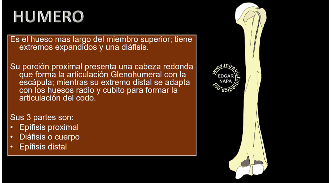
Su extremo proximal presenta una cabeza redonda que forma la articulación glenohumeral con la escápula.

Su extremo distal se adapta con los huesos radio y cubito para formar la articulación del codo .
Sus 3 partes son las siguientes:
EPÍFISIS PROXIMAL
DIÁFISIS
EPIFISIS DISTAL

INSERCIONES MUSCULARES EN EL HUMERO


SUBESCAPULAR
REDONDO MENOR
SUPRAESPINOSO
INFRAESPINOSO
DELTOIDES
PECTORAL MAYOR
ANCHO DORSAL
REDONDO_MAYOR
CORACOBRAQUIAL
TRÍCEPS BRAQUIAL
BRAQUIAL ANTERIOR
FLEXOR SUPERFICIAL DE LOS DEDOS
FLEXOR RADIAL DEL CARPO
PALMAR_LARGO
PRONADOR REDONDO
FLEXOR CUBITAL DEL CARPO
EXTENSOR DE LOS DEDOS
EXTENSOR DEL MEÑIQUE
ANCÓNEO
BRAQUIORRADIAL
SUPINADOR
EXTENSOR RADIAL LARGO DEL CARPO
EXTENSOR RADIAL CORTO DEL CARPO
EXTENSOR CUBITAL DEL CARPO


INSERCIONES MUSCULARES EN EL HUMERO


TAMBIÉN TE PUEDE INTERESAR

CUBITO

RADIO

ESCAPULA

ARTERIA BRAQUIAL

NERVIO RADIAL

information

This is an excellent place for you to add a paragraph and let your visitors know more about you and your services. Easily edit by go to tab pages and content and click edit.

INICIO | Anatomía humana | Anatomía Imagenológica | Semiología imagenológica | Diagnostico por imagenes

Contact Us

  • Your address
  • admin@admin.com

Follow us

2025 Company Inc. All rights reserved. Powered by RVsitebuilder
  • INICIO
  • Anatomía humana
  • Anatomía Imagenológica
  • Semiología imagenológica
  • Diagnostico por imagenes