• Login
  • INICIO
  • Anatomía humana
  • Anatomía Imagenológica
  • Semiología imagenológica
  • Diagnostico por imagenes

  • Descripción de diversos temas de anatomía humana, imagenología y diagnóstico por imágenes, apoyado por esquemas, dibujos y Gifs para su mejor compresión.


    • Anatomía humana .
    • Anatomía imagenológica .
    • Diagnóstico por imágenes .


FASCIAS DEL CUELLO.
I.  CERVICAL  SUPERFICIAL :  Cubre al músculo platisma.

II. CERVICAL PROFUNDA 
A. HOJA SUPERFICIAL (hoja muscular mayor). Es de REVESTIMIENTO. Envuelve a los músculos Esternocleidomastoideo y Trapecio . 
B. HOJA MEDIA (hoja muscular menor). Envuelve a los  músculos infrahioideo 
C. HOJA PROFUNDA (Pre-vertebral). Envuelve los  músculos largo del cuello, recto anterior, recto lateral de la cabeza . 
D. HOJA VISCERAL . Cubre a la Laringe , Tráquea , Faringe , Glándula Tiroides .     
E. HOJA VASCULAR (vaina carotidea). Esta formada por las hojas anteriores. Envuelve al nervio neumogástrico (X) , arteria Carótida común , vena Yugular interna    
ESPACIO PERIVERTEBRAL

COMPARTIMIENTOS PREVERTEBRAL Y PARAESPINAL
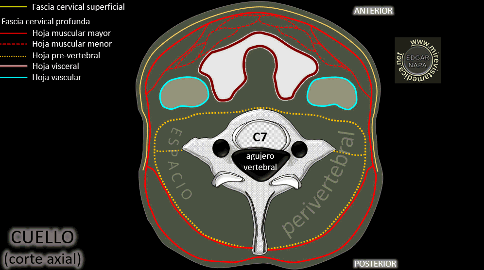
HOJA VASCULAR

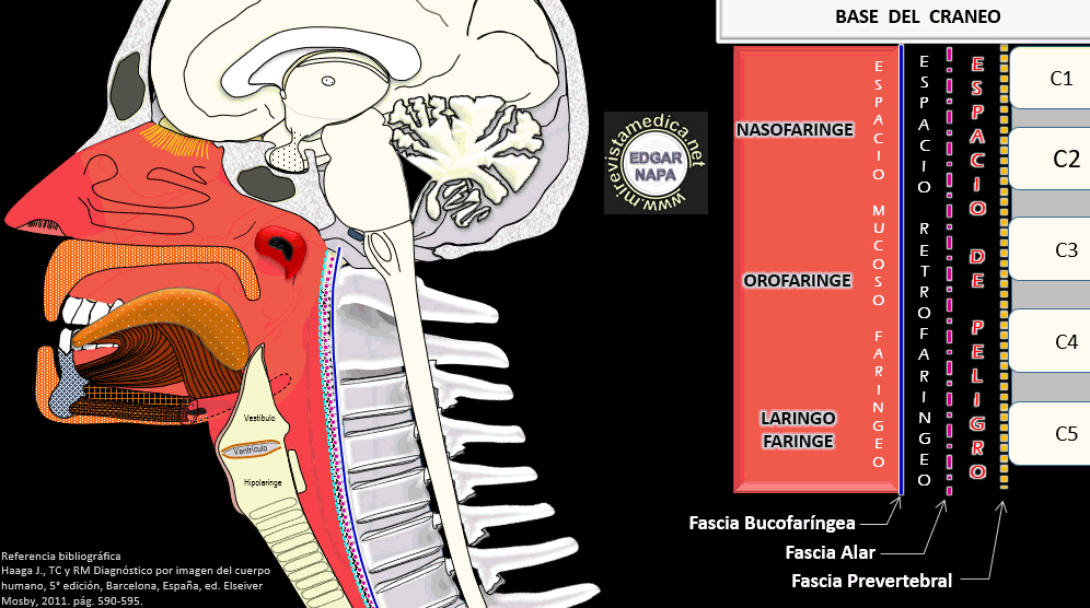
ESPACIOS MUCOSO FARINGEO, RETROFARINGEO Y DE PELIGRO

Triángulos del cuello

Drenaje venoso del cuello

Plexo cervical

information

This is an excellent place for you to add a paragraph and let your visitors know more about you and your services. Easily edit by go to tab pages and content and click edit.

INICIO | Anatomía humana | Anatomía Imagenológica | Semiología imagenológica | Diagnostico por imagenes

Contact Us

  • Your address
  • admin@admin.com

Follow us

2025 Company Inc. All rights reserved. Powered by RVsitebuilder
  • INICIO
  • Anatomía humana
  • Anatomía Imagenológica
  • Semiología imagenológica
  • Diagnostico por imagenes